Bone densitometry, also known as dual-energy X-ray absorptiometry (DXA or DEXA) scan, is a diagnostic test used to measure bone mineral density (BMD).
- Purpose:
- Procedure:
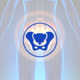
- Measurement sites:
- DXA T-score:
- Importance of bone density testing:
- Preparation and safety:
- Result:
Bone densitometry is primarily performed to assess the strength and density of bones, aiding in the diagnosis and monitoring of osteoporosis or other conditions that affect bone health.
During a bone densitometry scan, the patient lies on a padded table while a scanner arm passes over the body, emitting low-dose X-rays. The machine measures the amount of X-ray energy absorbed by the bones, which helps determine their density and strength.
Common areas assessed by bone densitometry include the spine, hip, and sometimes the forearm. These regions are most susceptible to fractures associated with osteoporosis.
The results of a bone densitometry scan are reported as T-scores, which compare the patient’s bone density to that of a healthy young adult population.
Bone densitometry is a crucial tool for assessing fracture risk, guiding treatment decisions, and monitoring response to therapy in individuals at risk of osteoporosis or other bone-related conditions.
Bone densitometry is a non-invasive and painless procedure that usually requires no special preparation. The amount of radiation exposure during a DXA scan is minimal, making it safe for most individuals.
The results of a bone densitometry scan should be discussed with a healthcare professional who can interpret the findings in the context of your medical history, overall health, and other risk factors for bone-related conditions. They can provide appropriate recommendations for treatment, lifestyle modifications, and monitoring.
BMD at Harley Street
At Harley Street Radiology Department, we are equipped with state-of-the-art imaging technology called Stratos DR –3D DXA (Dual-Energy X-ray Absorptiometry), an advanced imaging technology for assessing bone health.
3D-DXA is a breakthrough technology that uses routine BMD images to modelize a 3D image of the femur. Unlike traditional DXA scans, which provide 2D images, 3D DXA creates a three-dimensional representation of the bones, offering enhanced visualization and analysis. 3D-DXA provides automated workflow, retrospective analysis, patient follow-up and Report generation.


 أنقر هنا
 أنقر هنا